

Health & Beauty
WHAT ARE THE MAIN REGIONS OF THE SPINE?
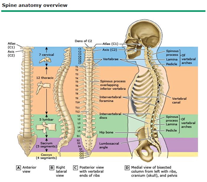
When most people talk about a back problem, they are actually referring to the spine in the vast majority of cases. The spine extends from the base of the skull down the back to the pelvis. It is made up of 33 skeletal pieces called vertebrae, each about 1.5 cm thick and stacked one on top of the other.
ON THIS PAGE
How many parts does the spine consist of?
What is an intervertebral disc?
What are facet joints?
What is a vertebral body?
How can the spine be flexible and yet resistant to movement?
Where do spinal nerves go after they leave the spine?
What about the spinal cord?
HOW MANY PARTS IS THE SPINE MADE UP OF?
Although the spine is a continuous structure, it is often described as having five different sections:

Cervical – the neck and upper back, consisting of 7 vertebrae that are adjacent to the skull. It supports the weight and movements of the head and protects the nerves that leave the skull.
Thoracic – the middle back, consisting of 12 vertebrae located between the cervical and lumbar spine
Lumbar – the lower back, consisting of 5 vertebrae, supports almost all of your body weight.
Sacrum – the base of the spine that is composed of 5 vertebrae fused (joined together) as a solid body, it attaches to the iliac bones of the pelvis to form the sacroiliac joints.
Coccyx – located below the sacrum, consisting of 4 fused vertebrae.
WHAT IS AN INTERVERTEBRAL DISC?
An intervertebral disc (or intervertebral fibrocartilage) is a soft cushion located between adjacent vertebrae of the spine. There are 23 intervertebral discs in the human spine, allowing smooth movement of the vertebrae, acting as a ligament that holds the vertebrae together, and functioning as a shock absorber for the spine. Located between 2 vertebrae, the intervertebral disc functions as a shock absorber between two bony structures. The disc has 2 components:
The annulus fibrosus – the tough outer portion.
The nucleus pulposus – the gelatinous central portion.
WHERE DO SPINAL NERVES GO AFTER THEY LEAVE THE SPINAL CORD?
The spinal canal and intervertebral foramina in the lumbar spine are bony tunnels that the spinal nerves travel through on their way to the periphery. When the diameter of these tunnels is reduced, there is less space for the nerve structures and, as a result, pressure can occur at this level.
WHAT ARE THE FACETS (PROCESSES) OF THE JOINT?
There are 2 pairs of protrusions through which the vertebrae connect to each other, including:
Superior articular facets, oriented superiorly
Inferior articular facets, oriented inferiorly
The points of connection between two vertebrae are the articular facets that keep the spine aligned during movement. Similar to other joints, the articular facets are aligned with the help of a membrane called synovium, which produces a fluid that allows one to slide over the other.
WHAT IS THE VERTEBRAL LAMA?
The bony structure located behind the spinal canal, in which the spinal cord is located. The vertebral laminae meet in the midline, from which the spinous processes (bony protrusions that can be felt through the skin in the middle of the back) develop. These laminae are often resected (laminectomy) to decompress the nerves in the spinal canal.
WHAT CAN BE SAID ABOUT THE SPINAL CORD?
The spinal cord descends through the center of the spinal canal, as a bundle of cells and nerve fibers that transmit electrical signals both to and from the brain, and to and from the rest of the body through the 31 pairs of spinal nerves that leave the spinal canal through the space between the vertebrae.
The spinal cord ends in the upper portion of the lumbar segment (at the L1 or L2 vertebral body). The last portion of the spinal cord is known as the “cone” /”conus medullaris”. Pressure on the conus medullaris can cause problems with sphincter control (for urine and feces) and numbness around the anus and genitals (an aspect called “hypoesthesia in SA”).
Below the terminal portion of the spinal cord, the spinal canal is occupied by the spinal nerves (grouped under the name of “horsetail”, after the appearance it imitates).
HOW CAN THE SPINE BE ELASTIC BUT ALSO RESISTANT TO MOBILIZATION?
Supporting the spine and providing, at the same time, flexibility – are the ligaments and muscles (resistant bands of connective tissue that insert and keep the bones attached)
Two main ligaments:
The anterior longitudinal ligament.
The posterior longitudinal ligament.
Both run along the entire spine, holding all its components together.
Source: danmartin.ro